Wednesday, August 31, 2011
Character head construction
Tuesday, August 23, 2011
Medical Transcription-Anatomy and Physiology of the Skin
Introduction to skin
Human skin is considered to be the largest organ of the body. The surface area of the skin on an average adult is 1.8 m2, and represents 16% of the total body weight. The thickness of the skin varies throughout the body. It depends on how much use we make of that area. For example, because we use our feet for walking, it is thickest on the soles of our feet. We use our hands for doing many everyday tasks such as picking up things and writing, so it is also thick on our palms.
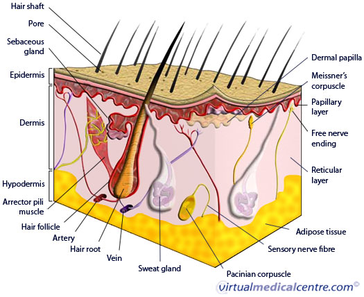
The skin is a multifunctional organ. It is divided into two main layers, the dermis and epidermis. The image shows a microscopic cross-section of human skin.
What does skin do?
Thermoregulation
The skin helps us to maintain our body temperature. When we are hot, there is vasodilation (widening of blood vessels) at the skin surface. This cools us down by allowing more heat to escape. When we are cold, there is constriction (narrowing of blood vessels). This allows less heat to escape, helping conserve heat.
Metabolism
When we are hot or exercising, sweat glands in our skin excrete water salts and proteins. Once on the surface of the skin, sweat evaporates into the air. This cools the skin and helps us control our body temperature.
Sensation
There are many nerve endings and receptors that sense changes in the skin. This allows us to feel everyday objects, feel pain, determine hot from cold and also sense pressure.
Protection
As the skin covers our whole body and is a continuous layer, it acts as a barrier and protects the body from injury and infection. It also shields against the sun's light and radiation and prevents us from drying up.
Synthesis of vitamin D
When exposed to the suns rays, the skin produces vitamin D3. This is essential for building strong, well shaped bones.
The epidermis
 The epidermis is the outermost layer of the skin. This layer consists of many special cells, including keratinocytes and melanocytes. Keratinocytes are cells that make a special fat which gives skin it's waterproof properties. Melanocytes produce melanin, which is a pigment giving us the colour of our skin. This layer is continuously shed and replaced, every 15–30 days.
The epidermis is sub-divided into 5 layers.
Stratum corneum
The outermost layer of the epidermis. There are many cells which are tightly packed together, This allows the skin to be tough and waterproof. This layer is important in the prevention of invasion from foreign things, such as bugs and bacteria.
Stratum lucidum
This layer contains several clear and flat dead cells. It is a tough layer and is found in thickened skin, including the palms of the hand and soles of the feet.
Stratum granulosum
The stratum granulosum is composed of 3 to 4 layers of cells. Here, keratin is formed, which is a colourless protein important for skin strength.
Stratum spinosum
This layer contains cells that change shape from columnar to polygonal. Keratin is also produced here.
Stratum basale
This layer is the deepest layer of the epidermis, in which many cells are active and dividing. The stratum basale is separated from the next layer – the dermis – by a basement membrane, which is a layer made of collagen and proteins.
The dermis
The dermis is the second major layer of the skin. It is a thick layer made up of strong connective tissues. It is further divided into two levels – the upper is made of loose connective tissue, called the papillary region, and the lower layer is made of tissue that is more closely packed, called the reticular layer. The dermis is made up of a matrix of collagen, elastin and network of capillaries and nerves. The collagen gives the skin its strength, the elastin maintains its elasticity and the capillary network supplies nutrients to the different layers of the skin. The dermis also contains a number of specialised cells and structures.
These include: hair follicles, sweat glands, sebaceous glands (produce sebum which helps lubricate skin & hair) and nails.
It also plays an important part in controlling our skin temperature and acts as a cushion against mechanical injury. When injured the dermis heals through the formation of granulation tissue (a tissue rich in new blood vessels and many different cells). This tissue helps pull the edges of a cut or wound back together. It takes our body from 3 days to 3 weeks to form this tissue.

Reference
- Ross MH, Kaye GI, Pawlina W. Histology: A text and atlas (4th edition). USA: Lippincott Williams & Wilkins; 2003.
- Saladin KS. Anatomy and Physiology: The unity of form and function (3rd edition). USA: McGraw Hill; 2004.
- Kumar P, Clark M. Clinical Medicine (5th edition). United Kingdom: WB Saunders; 2002.
- Revis DR, Seagle MB. Skin anatomy [online]. E-medicine; 2006 [cited 7 March 2006]. Available from URL:http://www.emedicine.com/plastic/topic389.htm
- Moore KL, Dalley AF. Clinically Orientated Anatomy (4th edition). Canada: Lippincott Williams & Wilkins; 1999.
- Kumar V, Abbas AK, Fausto N, Robbins SL, Cotran RS. Robbins and Cotran Pathologic Basis of Disease (7th edition). China: Elseiver Saunders; 2005.